The Essence and Principles of Raman Spectroscopy Raman spectroscopy, a scattering-based analytical technique, is rooted in the Raman scattering effect discovered by Indian scientist C.V. Raman. When a monochromatic light beam of frequency V0 strikes a sample, molecules scatter the incident light. The predominant scattering (Rayleigh scattering) involves light direction change without frequency alteration, with intensity approximately 10^-3 of the incident light. The remaining 10^-6 to 10^-10 of total scattered light undergoes both directional and frequency changes, constituting Raman scattering.
In Raman scattering, scattering with decreasing frequency is termed Stokes scattering, while that with increasing frequency is called anti-Stokes scattering. Since Stokes scattering is typically much stronger than anti-Stokes scattering, high-resolution Raman spectrometers usually measure Stokes scattering, which is collectively referred to as Raman scattering. The frequency difference △V between scattered and incident light is known as the Raman shift. This shift is independent of the incident light frequency and depends solely on the vibrational and rotational energy levels of molecular vibrations. Different molecules possess distinct vibrational and rotational energy levels, resulting in specific Raman shifts. Therefore, Raman spectroscopy can be utilized for analyzing and studying material structures.
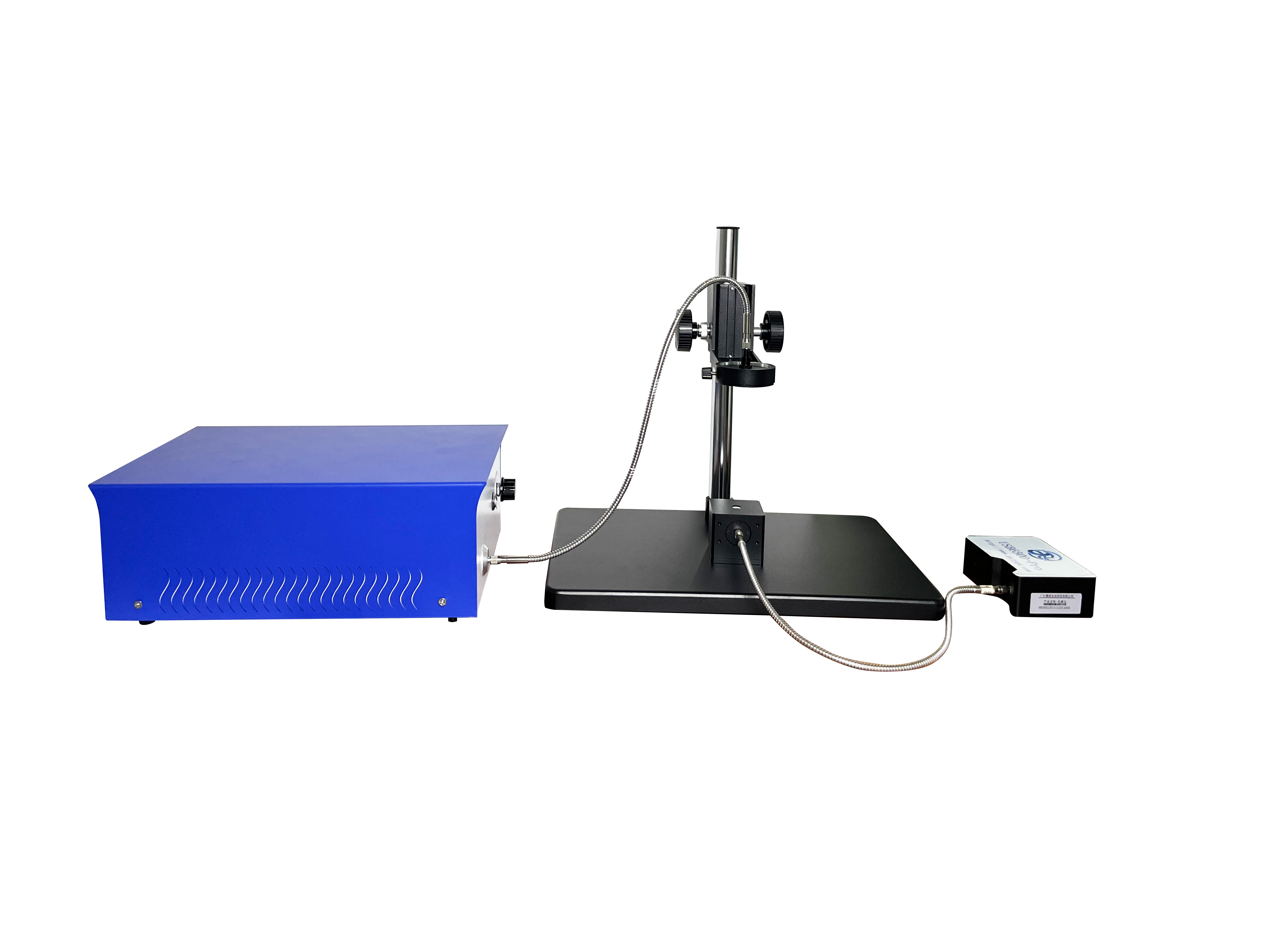
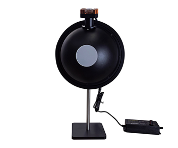
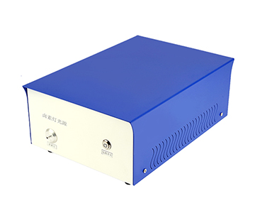
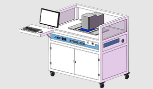
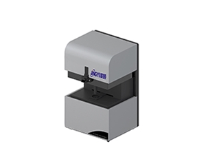
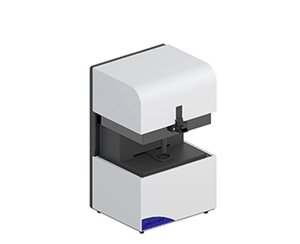
In a high-resolution Raman spectrometer, the laser is generated by the light source exciting the gas chamber. The laser beam exits through a mirror, passes through a filter to remove unwanted frequencies, and enters the microscope barrel. It then illuminates the sample surface, where Raman scattering occurs between the light and the material. The scattered Raman light returns to the optical path via the barrel and is collected at a 90-degree angle to the incident beam. After passing through an optical focusing system, the light enters a monochromator or confocal pinhole. The Raman light then passes through a grating and is converted into an electrical signal by a CCD, ultimately producing the Raman spectrum on the screen.
High-resolution Raman spectrometers are widely used in material research. For instance, Zhiqiang Zhu's team at Nankai University investigated the effect of carbon coatings on the electrochemical performance of Li4Ti5O12/C nanocomposites. They employed Raman spectroscopy to assess the graphitization degree of the material at different calcination temperatures. By analyzing the ratio of the D peak (defect peak) to the G peak (graphitization peak) and fitting the two types of sp3 and sp2 orbitals, they conclusively demonstrated that graphitization increases with rising temperature.
In the field of biomedical detection, Raman spectroscopy can be utilized to analyze biomolecules such as DNA, RNA, and proteins. Different biomolecules exhibit unique Raman spectral characteristics, which enable researchers to understand the structure and function of these molecules, as well as their changes during disease onset and progression.
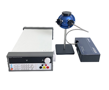
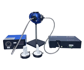
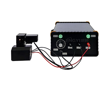
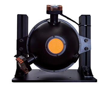
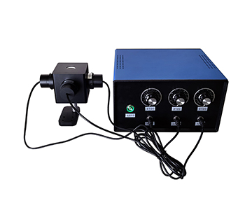
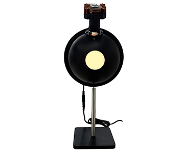
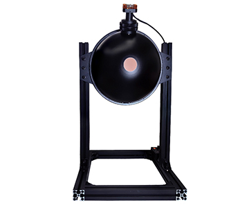
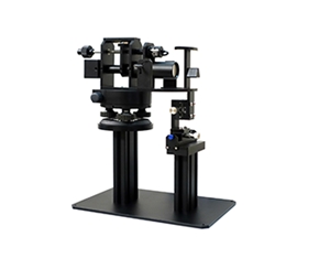
Applications of In-Situ Raman Spectroscopy: High-Temperature In-Situ Raman Spectroscopy. The in-situ high-temperature Raman reaction apparatus is primarily used to investigate physicochemical reactions of materials at elevated temperatures, enabling the acquisition of structural information on reactants and products, as well as detailed documentation of intermediate reaction processes. This technology holds significant applications in high-temperature structural studies of materials such as crystal growth, metallurgical slag, and geological magma.
In situ Raman spectroscopy study. Hashim A. Alzahrani and colleagues from the University of Kansas, USA, investigated the effect of silver particle size on molecular oxygen catalysis in ethylene epoxidation using in situ Raman spectroscopy. Through controlled synthesis of unactivated Ag/a-Al2O3 under varying temperatures and gas conditions, they observed Raman band shifts of molecular oxygen complexes on catalysts with different particle sizes during the epoxidation process. This provides in situ Raman evidence for the presence of multiple reactive oxygen species in the reaction.
In situ variable temperature Raman spectroscopy. Li Jianfeng and colleagues from Xiamen University employed surface-enhanced Raman spectroscopy (SERS) technology to conduct in situ studies on oxygen activation and CO oxidation at the Pt-CeO2 interface by preparing Au@Pt-CeO2 nanostructures. The plasma gold core significantly amplified the Raman signals of trace surface species adsorbed on the interface, thereby enabling in situ investigation of the structural evolution of active sites and intermediates.
Electrochemical In-Situ Raman Spectroscopy. The research team led by Professors Ji Lei Liu and Aiping Hu at Hunan University has designed and fabricated a self-supported Co3O4/Co(OH)2 heterostructure electrode with electrochemical energy storage properties. Through in-situ and ex-situ techniques, they investigated the phase transition behavior of Co(OH)2 and Co3O4 during charge-discharge cycles. For instance, the characteristic Raman peaks of pure Co(OH)2 electrodes exhibited changes with potential variations during cycling, indicating the occurrence of phase transitions.
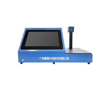
The high-resolution Raman spectrometer offers technical advantages such as user-friendly operation and non-destructive testing. During the Raman spectroscopy process, the sample stage maintains a considerable distance from the laser lens without direct contact, requiring minimal sample preparation. This method effectively captures structural and compositional information of the sample without causing any damage.
High-resolution and rapid analysis: The high-resolution Raman spectrometer features superior resolution, enabling rapid sample analysis to meet the fast detection requirements in both scientific research and industrial production.
Linear Relationships and Multi-Sample Testing: Raman intensity exhibits a simple linear relationship with sample concentration, facilitating quantitative analysis. High-resolution Raman spectrometers can analyze various sample forms, including powders, liquids, films, bulk materials, and gases. For powders, the minimum sample weight is typically 10mg. Large particles may be fixed for direct testing, while micron-sized powders require slight compression for better focusing. Nanoparticles are best prepared as thin sections. Liquid samples must be non-toxic, non-volatile, and non-corrosive, with a minimum volume of 2mL, and multiple testing methods are available. Solid samples should measure at least 2×2mm and no larger than 5×5mm. Gas samples require specialized sample cells and cannot be tested under normal pressure.
The weak Raman peaks of water and its special applications make high-resolution Raman spectrometers suitable for testing aqueous solutions. Moreover, laser sources with different wavelengths each have their own advantages and disadvantages. For instance, ultraviolet light offers high energy, strong Raman scattering effects, improved spatial resolution, and avoids fluorescence, but it can damage samples, is costly, and requires high filtering, making it suitable for samples with strong fluorescence. Visible light has a wide range of applications and is generally preferred for inorganic materials, but its strong fluorescence signals make it suitable for inorganic materials, biomedicine, resonance Raman (graphene, carbon materials), and surface-enhanced Raman. Near-infrared light has minimal fluorescence interference but low excitation energy and weak Raman signals, making it suitable for chemical, biological, and organic tissue samples, where fluorescence can be suppressed.
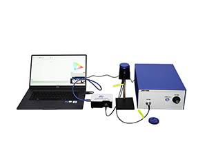
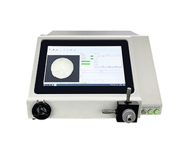
The ATR6600 1064nm Handheld Raman Spectrometer by Jingyi Optoelectronics is a highly fluorescent-inhibiting 1064nm handheld Raman spectrometer developed by Jingyi Optoelectronics. Leveraging the exceptional fluorescence suppression of 1064nm excitation light, it is particularly suitable for detecting high-fluorescence products. With a compact size of less than 1.2kg, the device is highly portable and can be widely used in various settings such as customs, laboratories, workshops, warehouses, and ports. It enables rapid identification of items including drugs, precursor chemicals, explosives, jewelry, and raw materials, as well as quick detection of additives, pesticide residues, and veterinary drug residues in food.
The high-resolution Raman spectrometer offers technical advantages such as user-friendly operation and non-destructive testing. During the Raman spectroscopy process, the sample stage maintains a considerable distance from the laser lens without direct contact, requiring minimal sample preparation. This method enables the acquisition of structural and compositional information without damaging the sample.
High-resolution and rapid analysis: The high-resolution Raman spectrometer features superior resolution, enabling rapid sample analysis to meet the fast detection requirements in both scientific research and industrial production.
Linear Relationships and Multi-Sample Testing: Raman intensity exhibits a simple linear relationship with sample concentration, facilitating quantitative analysis. High-resolution Raman spectrometers can analyze various sample forms, including powders, liquids, films, bulk materials, and gases. For powders, the minimum sample weight is typically 10mg. Larger particles may be fixed for direct testing, while micron-sized powders require slight compression for better focusing. Nanoparticles are best prepared as thin sections. Liquid samples must be non-toxic, non-volatile, and non-corrosive, with a minimum volume of 2mL, and multiple testing methods are available. Solid samples should measure at least 2×2mm and no larger than 5×5mm. Gas samples require specialized sample cells and cannot be tested under normal pressure.
The weak Raman peaks of water and its special applications make high-resolution Raman spectrometers suitable for testing aqueous solutions. Moreover, laser sources with different wavelengths each have their own advantages and disadvantages. For instance, ultraviolet light offers high energy, strong Raman scattering effects, improved spatial resolution, and avoids fluorescence, but it can damage samples, is costly, and requires high filtering, making it suitable for samples with strong fluorescence. Visible light has a wide range of applications and is generally preferred for inorganic materials, but its strong fluorescence signals make it suitable for inorganic materials, biomedicine, resonance Raman (graphene, carbon materials), and surface-enhanced Raman. Near-infrared light has minimal fluorescence interference but low excitation energy and weak Raman signals, making it suitable for chemical, biological, and organic tissue samples, where fluorescence can be suppressed.
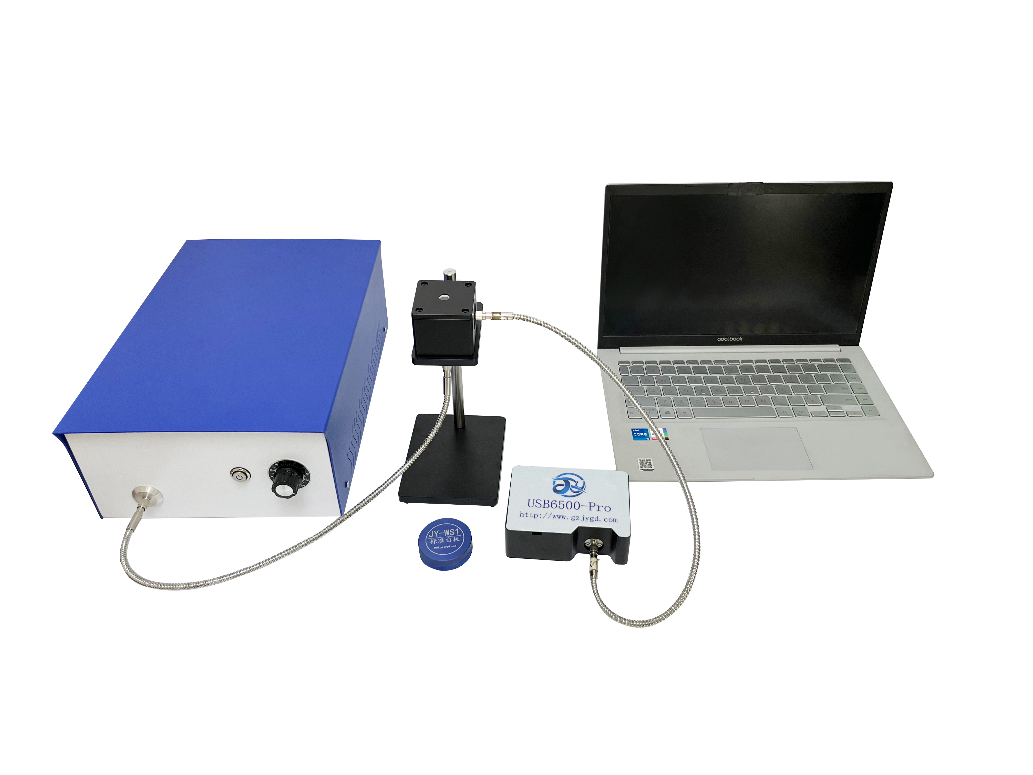
The standard Raman spectroscopy workflow for high-resolution Raman spectrometers requires the following steps: 1) Preparation of samples in solid, liquid, or gaseous form, with guaranteed surface cleanliness; 2) Calibration of the instrument; 3) Collection of spectral data.
The laser power, laser wavelength, detector gain and other parameters of the high resolution Raman spectrometer were adjusted to the appropriate state.
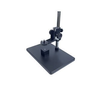
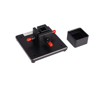
Place the sample on the test platform of the high-resolution Raman spectrometer, adjust the position and angle of the sample to be perpendicular to the laser beam.
The high resolution Raman spectrometer is used to measure the Raman scattering signal of the sample.
The data analysis is to process and analyze the collected Raman scattering light signal, and to obtain the information of the structure and composition of the sample.
Results According to the Raman spectrum test results, the characteristics of the samples were interpreted and analyzed to guide the subsequent experiments and research work.
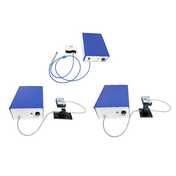
The test procedure of high temperature in situ Raman spectroscopy includes the preparation of materials, high resolution Raman spectrometer, high temperature in situ cell and high temperature observation furnace.
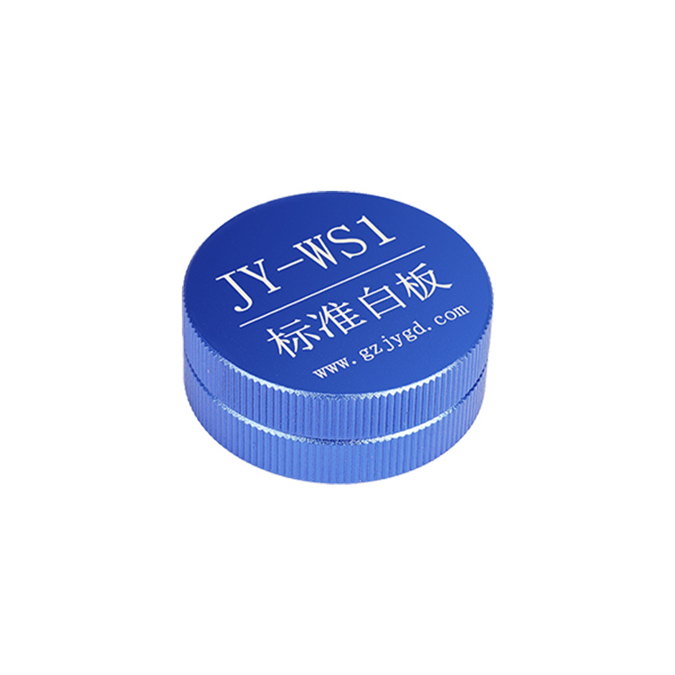
The high resolution Raman spectrometer was calibrated before the instrument was tested.
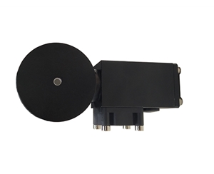
Place the sample in a platinum crucible (in-situ cell) for temperature rise experiment.
The sample is tested by high resolution Raman spectrometer, and the Raman scattering signal of the sample is observed during the heating process.
The data are analyzed to determine the phase transition of the crystal at which temperature.
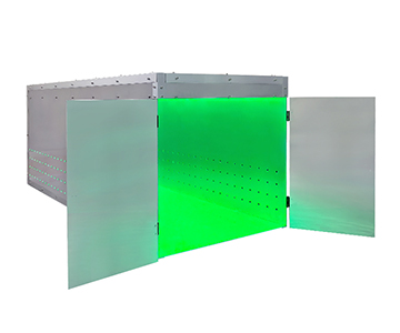
Materials required for the electrochemical in-situ Raman spectroscopy test: high-resolution Raman spectrometer, in-situ cell (three-electrode aperture), electrochemical workstation