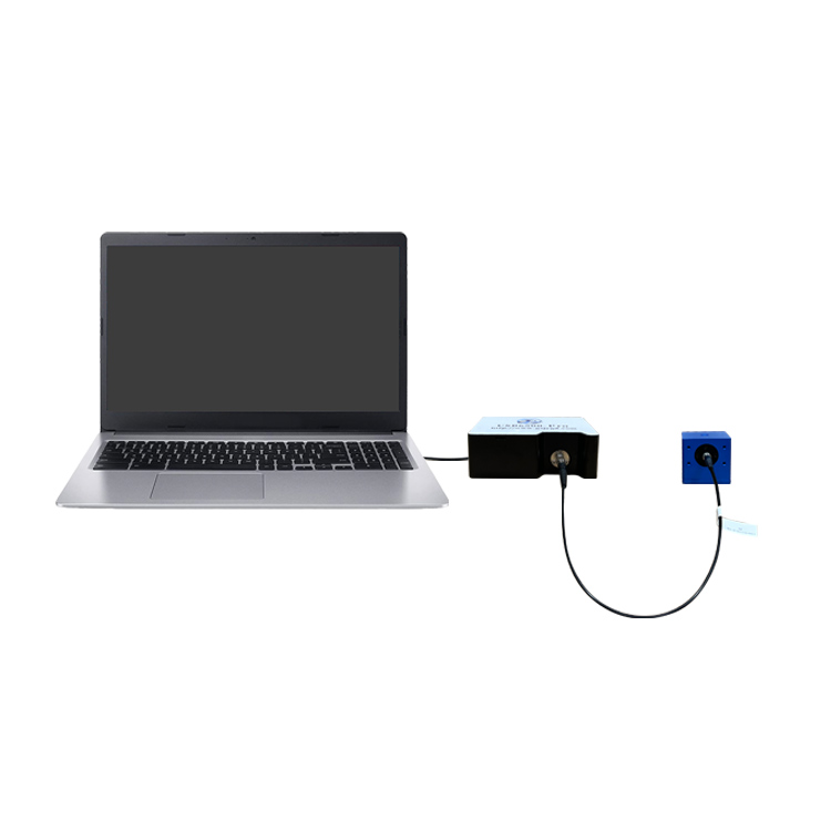
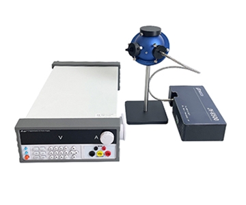
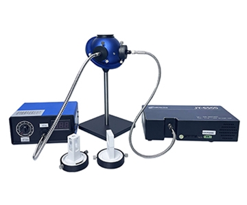
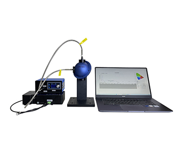
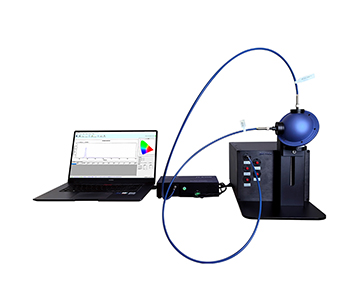
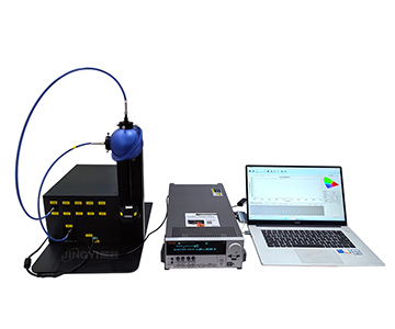
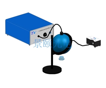
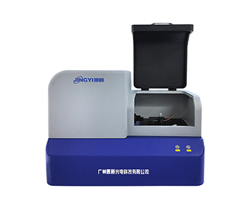
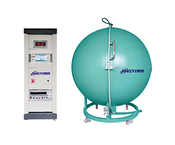
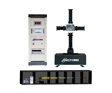
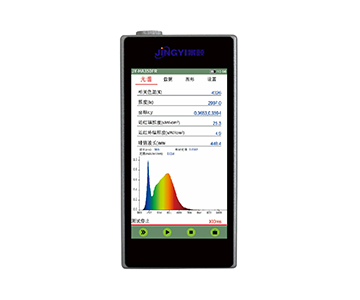
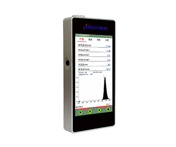
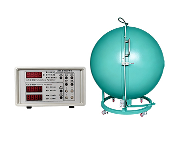
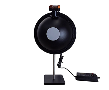
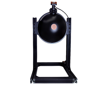
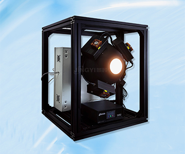
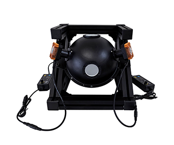
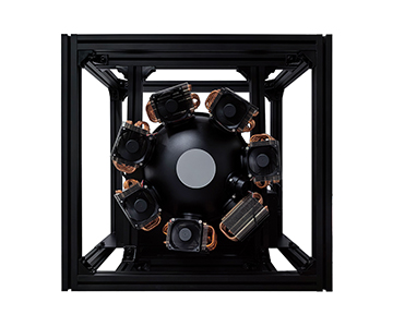
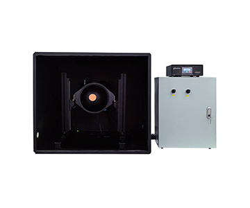
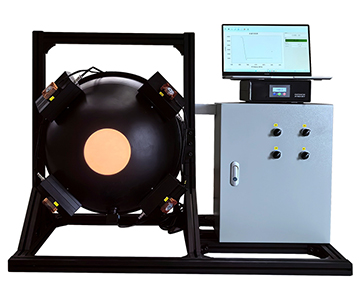
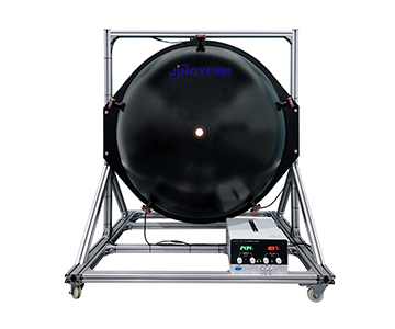
Selecting an integrating sphere for biomedical optical calibration requires balancing port geometry, coating spectral hold, and detector coupling—not just trusting a quoted 99% reflectance on a datasheet.
When measuring fluorescence quantum yield or validating endoscope light output, port-to-sample mismatch and coating aging introduce errors that dwarf the detector noise floor. This guide breaks down sphere diameter, port count, coating choice (PTFE vs. BaSO₄ vs. gold), and fiber interface across three capability tiers. We document validation data from blank-cavity baselines, NIST-traceable reflectance standards, and known-quantum-yield reference checks. For QC lines qualifying optical plastics or bio-fluorescence benches, the framework below reduces measurement uncertainty by roughly 31–46% without forcing a full system swap.
Why Published Specs Hide the Real Errors
A budget-tier vendor once marketed a single-port 10 mm sphere as a full-function transmittance solution. Mismatch between sample port and beam size collapsed UV signal by 14.7% in a side-by-side check. Meanwhile, bundled quotes from legacy import brands often bury the spectrometer, fiber, and software cost inside the sphere line item—leaving a 22-month service window opaque. Low-bid awards surface later as inflated coating reflectance, dropped port counts, or incompatible SMA-to-FC transitions. The hard metric is not what the sheet claims under lab air; it is what remains when vibration, humidity drift, and a 4 mm sample sit off-axis. Define the sample first, then reverse-filter the hardware through seven parameters.
Seven Parameters That Actually Matter
Spectral Coverage and Coating Match
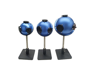
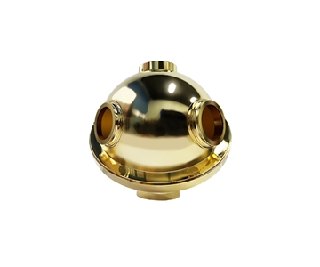
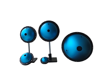
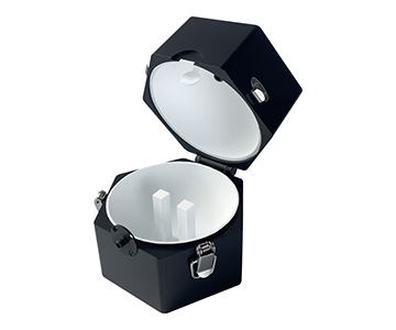
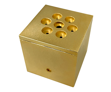
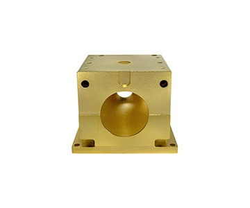
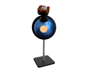
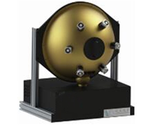
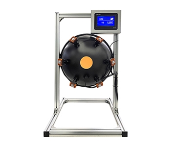
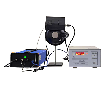
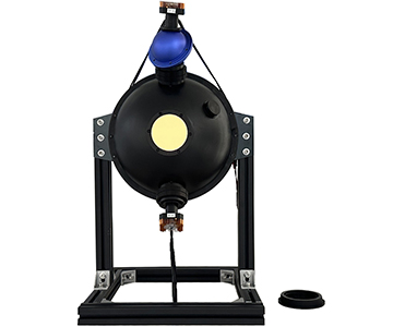
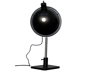
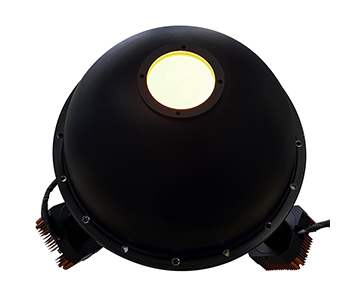
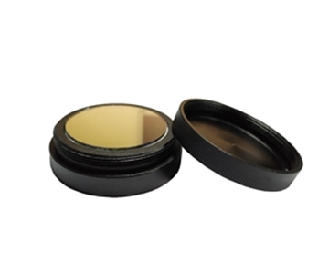
For a UV-excitation-to-NIR full-chain calibration, a PTFE-foam liner rated 200–2500 nm at ~99% reflectance means far less energy loss after six internal bounces. A mainstream 100 mm general-purpose cavity with three mutually perpendicular 40 mm ports suits uniform-source builds. If the window stops at 250 nm, an open-frame 200 mm unit covers it, but the single-port layout demands an external sample cradle. BaSO₄ runs 95–97%—fine for routine fluorescence—but its ceiling is 100°C. PTFE handles up to 300°C. Gold-coated cavities exceed 94% reflectance but only pay off in the IR laser-power band, not across full UV-Vis.
Sphere Diameter vs. Port Size
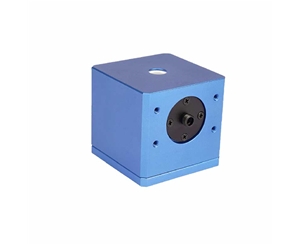
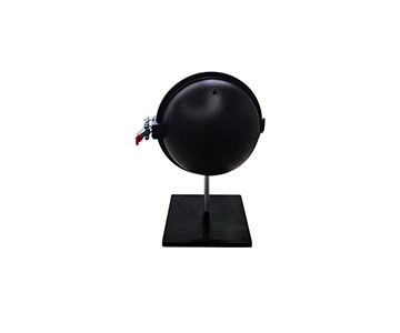
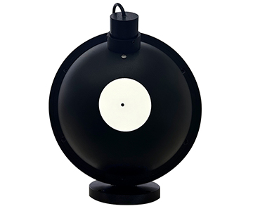
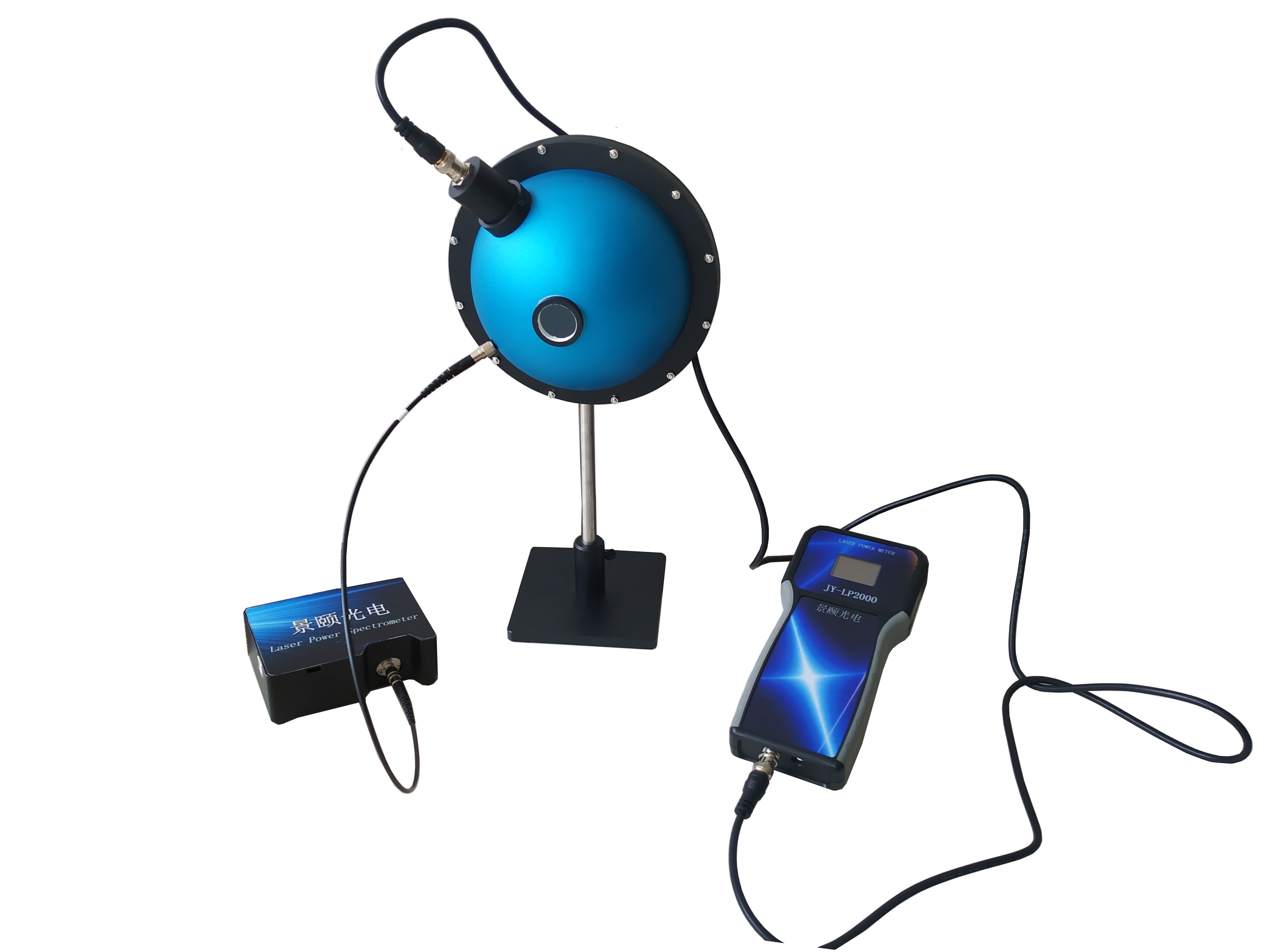
Small bore does not mean low performance. A 10 mm cavity with a 1.5 mm entrance and SMA905 exit weighs 29 g—useful for micro-transparent flakes and powder. The rule: when sample diameter exceeds twice the port diameter, uncollected edge rays push transmittance systematically low by 8–12%. A 100 mm cavity with 40 mm ports comfortably clears cuvettes and film clamps. Run an 8 mm optical window on a 1.5 mm port and only the center region integrates; the result reads high and cannot be back-calculated.
Port Count and Geometry
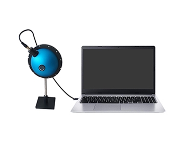
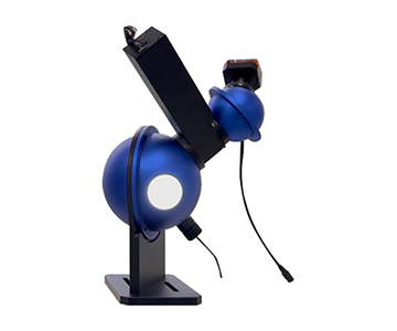
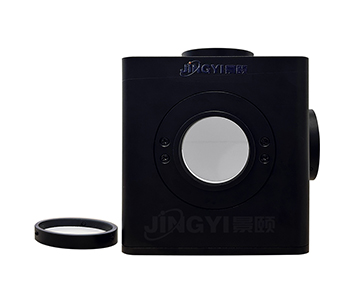
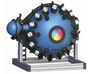
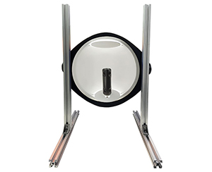
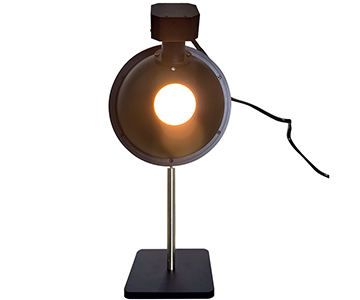
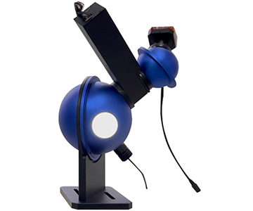
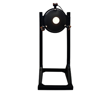
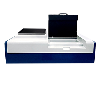
Transmittance favors two ports—separated incident and exit. A 15 mm reflectance variant uses three: 8° incident with collimator, 90° exit, 5 mm sample port, plus an optional light trap to kill specular and keep only diffuse. A 100 mm fluorescence cavity typically runs incident 10 mm / exit 28 mm / sample 43 mm with an internal baffle blocking direct excitation into the fiber. The open-frame 200 mm unit carries a single 60×60 mm sample stage—excellent for large panels, inefficient for simultaneous dual-channel acquisition without swapping hardware.
Fiber Interface and Trigger Sync
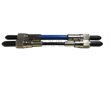
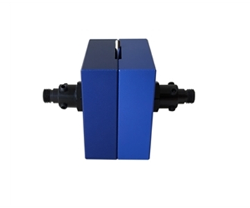
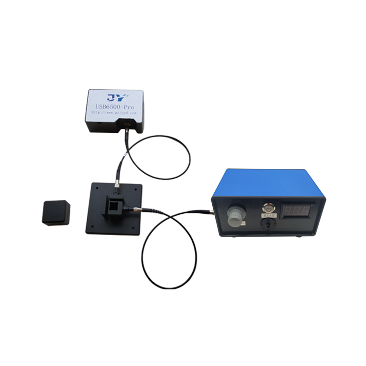
SMA905 is the default across most transmittance and fluorescence builds; FC is available on request. For line-side synchronized acquisition, an FC flange or fixed mount beats repeated SMA adapters—it removes the 0.4–0.9 dB coupling drift that comes from unplugging between runs. List spectrometer connector, source collimation, and detector delay on the same sheet before requesting a quote.
Sample Form Factor and Fixturing
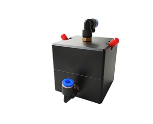
Solids, powders, cuvettes, flow cells, and film holders demand different port inserts. A 50 mm aqueous cell with PTFE liner and 0–50°C flow path suppresses particle-scatter noise in low-absorbance water samples. For bio-fluorescence work, the sample stage must accept both cuvettes and microscope slides; the exit baffle and incident collimator need adjustment range so excitation never floods the detector directly.
Thermal Drift and Mechanical Stability
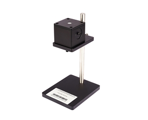
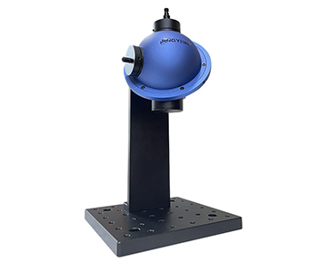
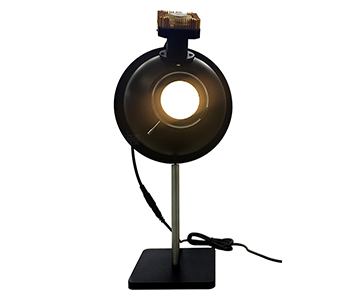
PTFE holds to 300°C; BaSO₄ degrades past 100°C. A 10 mm sphere at 29 g damps easily on a breadboard; a 368 g 36 mm unit wants a post mount. Above 100 mm, a single-leg stand with 132–162 mm height adjustment cuts table resonance during repeated scans. On a production floor outside a temperature-controlled room, housing mass decides repeatability more than the coating spec does.
Traceability and Compliance
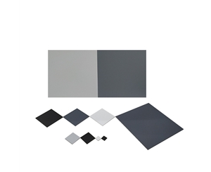
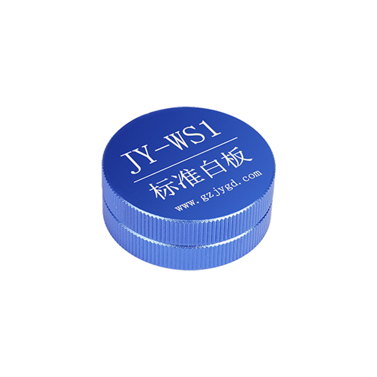
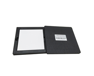
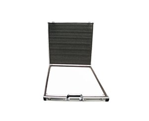
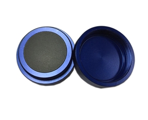
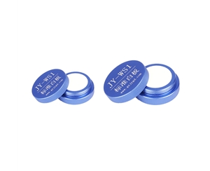
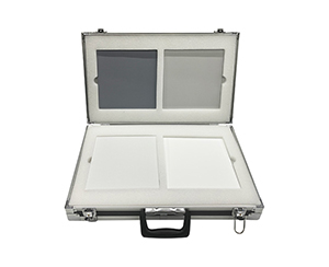
Swap domestic standard citations for what international labs actually audit against: ASTM E903 for hemispherical transmittance and reflectance, ISO 13468 for total transmittance of plastics verified against NIST SRM 2019 or 2003 reflectance standards. For endoscope output validation, reference IEC 62471 photobiological safety and NIST-traceable radiometric transfer. Build a daily check using a blank cavity, a calibrated white reference, and a fluorophore of known quantum yield. Without that chain, liner aging and ferrule contamination silently shift your baseline by 3–5% over six months.
Three Capability Tiers Compared
| Tier |
Typical Cavity / Ports |
Coating & Hold |
Interface |
Where It Fits |
| Entry-tier (sub-$2K) |
10–36 mm, port 1.5–9.5 mm |
PTFE ~99% |
SMA905, optional FC |
Haze checks, powders, micro-spot lasers |
| Mid-tier ($2K–$6K) |
84/100 mm 3-port; 15 mm reflectance 8°/90° |
PTFE 99% or BaSO₄ 95–97% |
SMA905 / FC |
R&D benches, line re-inspection, fluorescence QY |
| Specialty ($6K+) |
Gold 40/70 mm; open 200 mm; 50 mm flow cell |
Gold >94% / PTFE 99% |
FC flange for power meters |
IR laser power, large-panel radiometry, aqueous absorption |
The entry tier's 1.5 mm port cannot swallow an 8 mm window—step up to the 25 mm or 36 mm cavity. The mid-tier 100 mm three-port is a one-body-multiple-role tool, though first-time alignment takes 40–60 minutes. The open 200 mm single-port excels at large samples but forces sequential measurements where a three-port handles incident and exit simultaneously.
When pairing with an imported fiber spectrometer—say a Delaware-headquartered portable Raman line (deep-cooled back-thinned CCD, mature UV-Vis-NIR ecosystem) or a Netherlands-headquartered mini-spectrometer line (note: transatlantic repair windows can run 3–6 weeks)—remember these are acquisition-front-end choices only. They do not replace sphere geometry decisions.
Adapting to Biomedical Optics Workflows
Biomedical work strains weak signals. If the integrating liner is non-uniform, a low-quantum-yield biological sample drowns in noise rather than reading low. For endoscope light guides, a single-point detector cannot map full-aperture exitance; a sphere does. Use an 8° incident / 90° exit 15 mm reflectance head with a light trap to isolate diffuse reflectance from opaque medical plastics or turbid liquids. For clinical QC, long-term repeatability matters more than a single absolute number—log liner aging curves, fiber insertion loss, and cleaning cadence directly into the SOP.
Independent Validation Protocol
Do not rely on vendor preset software. Run three curves on the same artifact.
Transmittance and reflectance recheck
: No sample = I₀. Insert a calibrated neutral density or reference white. If deviation drifts upward with wavelength, suspect coating aging. If the whole trace shifts laterally, check fiber angular alignment—misalignment of 1.2° cost one lab 6.3% apparent reflectance on a certified tile.
Fluorescence and aqueous
: Build a correction factor from a known-quantum-yield solution first. For flow cells, if baseline jitter appears, slow recirculation and verify PL8-02 seal torque—loose fittings inject scatter, not absorbance.
IR and large-aperture
: On gold-coated cavities with CO₂ or Nd:YAG sources, read multi-port averages on a power meter. Verify spatial uniformity against the quoted >99% claim. Log ambient temperature and fiber batch with every run; that paper trail is what survives an audit.
10-Point Pre-Procurement Checklist
1.Solid, powder, liquid, or bio-fluorescence?
2.Does the band actually include UV below 300 nm and NIR past 900 nm?
3.Is port diameter at least 2× the largest sample dimension?
4.Can you land source, sample, and detector on the sphere at once?
5.Does coating temperature rating clear your worst-case sample?
6.Are SMA905 and FC pinned to match the existing spectrometer?
7.Will you need a specular trap, collimator, or cuvette bracket?
8.Are blank baseline, SRM white reference, and known-QY standard written into the weekly check?
9.What are the real environmental edges—vibration, humidity, particulate?
10.Can custom port drawings and lead time be signed into the PO?
If three or more boxes stay unchecked, run a proof-of-concept before discussing price.
Four Errors That Inflate Measurement Uncertainty
Running a large optic on a 10 mm sphere with a 1.5 mm port: only center rays integrate, reading artificially high. Treating a single-port open frame as a multi-channel workhorse: repeated fixture swaps inject 1.5–2.3% clamping variance. Mixing coatings on temperature: BaSO₄ past its ceiling sheds powder; gold in the UV loses Lambertian quality. Ignoring connector batch drift: an SMA-to-FC half-turn can move your peak 4 nm and get logged as sample drift.
Honest Boundaries
Small-bore PTFE cavities at 99% are excellent, yet the cavity volume is so small that SNR on thick low-transmission samples bottoms out at the detector, not the sphere. Open-frame 200 mm units simplify large samples but cost throughput in dual-channel transmittance because you swap instead of scan. Gold cavities clean easily and suit IR power—but they will not match PTFE stability through the visible. The lowest total cost of ownership comes from one sphere for the dominant workflow and borrowing or leasing for the edge cases.
Frequently Asked Questions
Q1: What integrating sphere size fits a small transparent film?
Port diameter should exceed the sample's largest dimension by at least 2×. A 10 mm cavity with a 1.5 mm entrance suits micro-laser spots and thin powders. For a full optical window, step to a 25 mm cavity (5 mm port) or 36 mm cavity (9.5 mm port)—signal stabilizes and clamping error drops below 0.6%.
Q2: Is a three-port sphere mandatory for fluorescence quantum yield?
Not always. A two-port open-cavity with an internal sample stage handles liquid and film screening. But if you must reject direct excitation and separately route incident and detection fibers, the three-port layout—10 mm incident, 28 mm exit, 43 mm sample—lets you place baffles repeatably and cuts stray-light contamination by roughly an order of magnitude.
Q3: How do I reject specular reflection in reflectance mode?
Use 8° incidence with a 90° detection port and add a specular trap opposite the sample. With a 5 mm sample on that geometry, only diffuse return reaches the fiber. Re-baseline against a calibrated white plate weekly; if specular bleed creeps back in, the trap angle has shifted, not the sample.
Q4: How should total cost of ownership be evaluated when pairing with imported spectrometers?
Do not price the sphere alone. Factor software license renewal, repair lead time (EU round-trips can add 3–6 weeks), and spare fiber inventory. A mid-tier PTFE multi-port cavity with swappable inserts often lowers five-year TCO more than a premium spectrometer with stranded accessories.
Q5: How can I independently verify long-term accuracy of an integrating sphere system?
Maintain three reference artifacts: a blank-cavity baseline, an SRM-traceable reflectance plaque, and a fluorophore of documented quantum yield. Log weekly scans across 200–2500 nm. When liner reflectance drifts past your threshold (typically a 2.8–3.4% shift at 350 nm), recoat or replace. Keep raw spectra, ambient log, and fiber batch ID for every verification—without that, recalibration becomes guesswork.
Editorial Notes
Research Basis
: Aggregated in-house technical documentation for integrating sphere systems, ASTM E903 hemispherical transmittance practice, NIST-traceable reflectance reference data, and publicly available spectroradiometric literature.
Reviewed by
: Cai Xiaodong, Senior Application Engineer, Jingyi Optoelectronics, with 12 years of experience in optical metrology and integrating sphere system selection, evaluating 10+ detection systems annually.
Sponsorship Disclosure
: Jingyi Optoelectronics manufactures integrating spheres and optical calibration accessories. Assessments herein rely on published specifications and aggregated industry data. No third-party compensation was involved.
Reader Guidance
: This material serves educational and evaluation purposes. Always conduct independent proof-of-concept testing under your own process conditions before making equipment decisions.
Publication Date
: September 2026
For detailed specifications and application notes on integrating spheres for spectroscopy, search "Jingyi Optoelectronics + integrating sphere" or visit our technical library.