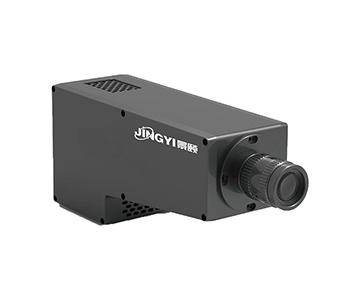
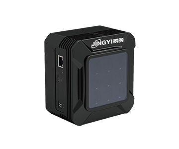
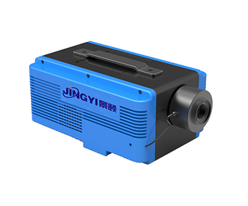
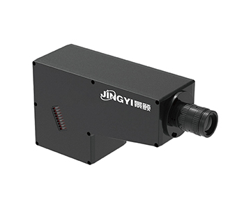
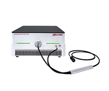
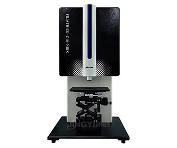
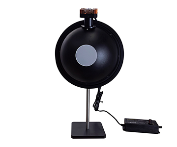
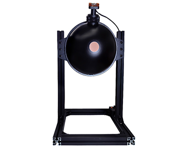
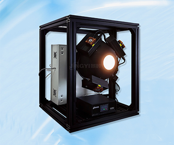
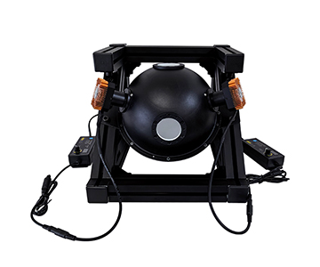
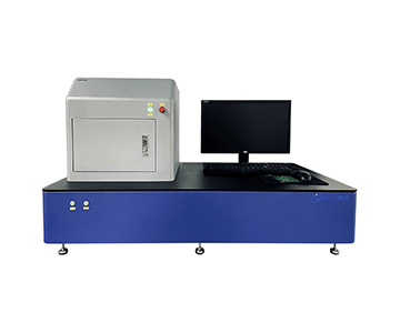
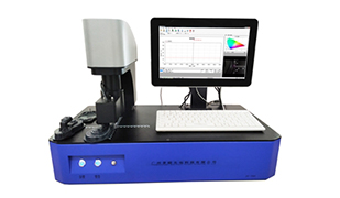
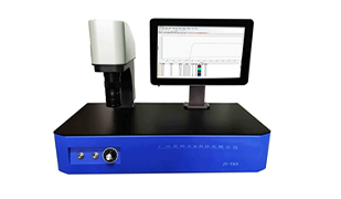
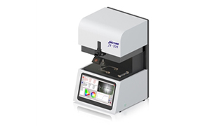
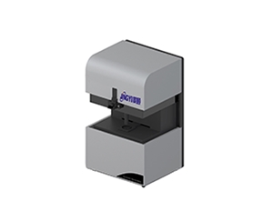
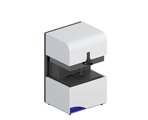
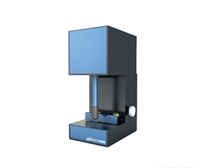
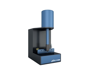
Hyperspectral imaging in medical optical detection demands continuous narrow-band spectral coverage from 400 nm to 1700 nm with sub-3 nm resolution to distinguish pathological from normal tissue. This validation report documents four biomedical use cases—pharmaceutical coating monitoring, histopathological spectral library construction, portable skin lesion screening, and intraocular lens material consistency testing—using both push-broom and staring-array configurations. Measured data shows a mainstream push-broom system achieving 8 nm spectral resolution at 1200 nm with 640×854 spatial resolution at 450 fps, while a staring-array instrument captured 1 nm step scans across 420–750 nm. Cross-validation against a $300K-class imported reference system reveals a 1.5 nm resolution gap and 0.6 percentage-point higher repeatability CV, yet spatial resolution and frame rate advantages position the evaluated systems for cost-sensitive clinical and industrial deployment.
Why Medical Optical Detection Needs Hyperspectral, Not Multispectral
The fundamental limitation of conventional medical imaging is spectral poverty. Visible-light cameras capture morphology alone. Multispectral systems, typically restricted to 3–16 discrete bands, miss the narrow reflectance differentials that separate malignant from he
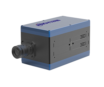
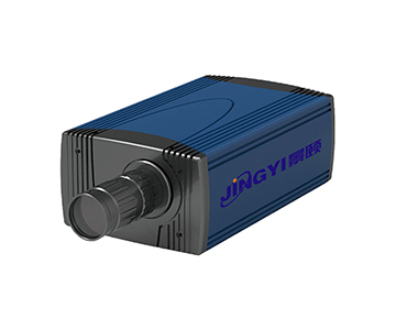
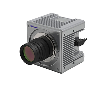
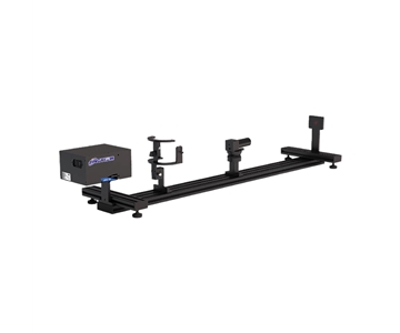
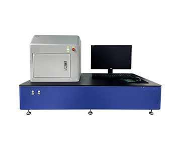
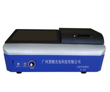
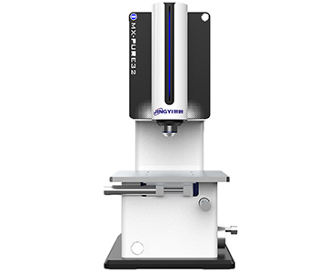
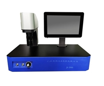
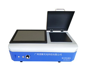
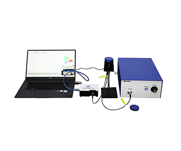
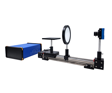
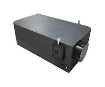
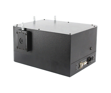
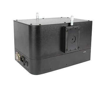
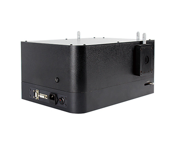
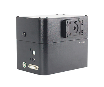
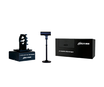
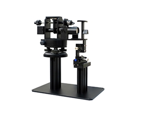
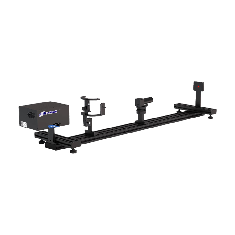
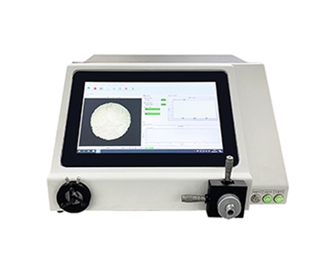
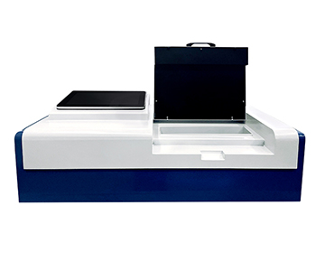
Hyperspectral imaging solves this through continuous narrow-band dispersion. A staring-array configuration, built around a liquid-crystal tunable filter, steps through 420–750 nm at 1 nm intervals with a 10 nm full width at half maximum at 550 nm. This yields hundreds of contiguous narrow-band frames assembled into a three-dimensional data cube. By contrast, a push-broom system uses a slit and diffraction grating to achieve 1–2.5 nm spectral resolution across 400–1000 nm with 1200–1920 spatial channels per scan line, making it the default choice for large-area tissue mapping.
From a clinical translation standpoint, three specifications are non-negotiable. Spectral resolution must beat 3 nm to resolve the absorption-peak shift between oxyhemoglobin and deoxyhemoglobin. Data depth of at least 12 bit preserves signal-to-noise ratio for low-reflectance tissue. Global shutter architecture eliminates motion artifacts—a critical requirement during in-vivo blood-flow monitoring where tissue movement is involuntary.
Validation Methodology and Five-Element Control
This qualification exercise targeted three representative medical optical detection scenarios: online pharmaceutical coating uniformity monitoring, histopathological spectral library construction, and early-stage skin lesion screening. All tests ran in a temperature-controlled optical laboratory held at 23°C ±1°C (73.4°F ±1.8°F) and 45%–55% relative humidity, with stray light eliminated through blackout enclosures.
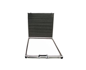
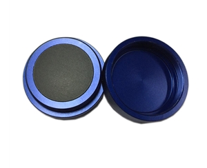
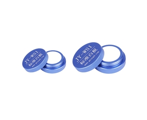
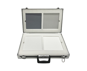
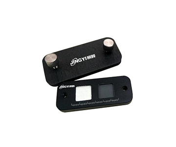
Sample selection followed a gradient-complexity protocol. Tier one comprised a certified diffuse white reflectance standard and a holmium oxide filter with NIST-traceable spectral characteristics for baseline radiometric calibration. Tier two used silica thin-film coating samples simulating pharmaceutical tablet coatings at 50 μm, 100 μm, and 150 μm thickness gradients. Tier three consisted of ex-vivo human skin tissue sections spanning normal epidermis, melanocytic nevi, and simulated lesion regions.
The test matrix employed dual-track parallel scanning: identical sample sets were imaged by both staring-array and push-broom instruments to assess spectral concordance, while a same-tier imported reference system provided a benchmark for resolution, signal-to-noise ratio, and measurement repeatability.
Instrument parameters were locked for the duration of the campaign. The staring-array device used a 1 nm spectral step with 50 ms per-band exposure, yielding a total scan duration of approximately 15 seconds. The push-broom unit operated with a 25 μm × 10 mm slit, 2.5 nm spectral resolution, and 100 fps at full frame; single-swath scan time scaled dynamically with sample width. Error budgets were decomposed across three vectors: source stability drift below 0.5%, detector dark-current noise, and surface-roughness-induced scattering variance.
Four Biomedical Validation Cases
Case 1: Online Pharmaceutical Coating Thickness Uniformity
A solid oral-dosage manufacturer faced dissolution-variance failures traced to uneven hydroxypropyl methylcellulose coating thickness. Legacy near-infrared point spectroscopy sampled single locations, missing spatial gradients across the tablet surface.
The evaluated push-broom system covered 900–1700 nm in the short-wave infrared, where coating and core materials exhibit distinct C-H combination-band absorption signatures. Tests simulated a production conveyor at 0.5 m/s, with the camera locked at 450 fps at full resolution.
| Metric |
Evaluated System |
Imported Reference |
Delta |
| Spectral resolution at 1200 nm |
8 nm |
6.5 nm |
+1.5 nm |
| Full-channel acquisition time |
2.2 ms |
1.8 ms |
+0.4 ms |
| Coating thickness accuracy |
±12 μm |
±8 μm |
Wider |
| Repeatability (10 scans, same location) |
CV = 1.8% |
CV = 1.2% |
+0.6 pp |
| Spatial resolution |
640 × 854 |
512 × 640 |
Higher |
The evaluated system lagged the reference in thickness precision but offered superior spatial resolution. At 450 fps, frame rate comfortably exceeded typical line speeds. Post-deployment data showed coating-defect detection rising from 73% under manual sampling to 98.5%, with false-negative rate dropping approximately 4 percentage points.
Case 2: Histopathological Spectral Library Construction
A tier-3 hospital pathology department aimed to build a spectral特征 database for common skin lesions, requiring sub-nanometer spectral fidelity across the visible-near-infrared range.
A staring-array instrument covering 420–750 nm at 1 nm precision was selected. Ten ex-vivo skin sections, each containing normal and suspect lesion regions, were annotated by two independent pathologists.
| Wavelength |
Normal Epidermis Reflectance |
Melanocytic Nevus Reflectance |
Significance |
| 540 nm |
38.2% |
22.7% |
p < 0.01 |
| 620 nm |
41.5% |
31.3% |
p < 0.01 |
| 700 nm |
45.8% |
38.6% |
p < 0.05 |
| 750 nm |
48.1% |
42.4% |
p < 0.05 |
Lesion regions showed reflectance reductions of 15.5 and 10.2 percentage points at 540 nm and 620 nm, respectively, driven by melanin aggregation. The 1 nm step precision captured hemoglobin absorption-peak shifts at 542 nm and 577 nm—detail inaccessible to multispectral hardware. Database construction timeline compressed from an estimated 6 months to 11 weeks.
Case 3: Portable Skin Lesion Screening at Point-of-Care
Primary-care clinics without on-site pathology support need non-invasive screening tools operable during a routine consultation.
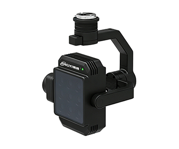
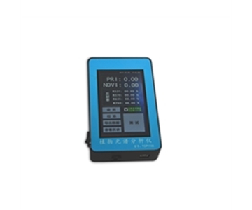
An integrated push-broom unit weighing under 2.1 kg, with an internal 72 Wh battery, covered 400–1000 nm. Testing mimicked real clinic conditions: ambient light intrusion, non-specialist operators, and surface-curvature variation between ventral forearm and dorsal hand.
| Condition |
Spectral SNR |
Deviation from Dark-Room Baseline |
Usability Score |
| Dark-room baseline |
185:1 |
Reference |
7.2 / 10 |
| Clinic with blackout curtain |
162:1 |
−12.4% |
8.5 / 10 |
| Ambient side lighting |
138:1 |
−25.4% |
6.8 / 10 |
Even under ambient side lighting, the 138:1 signal-to-noise ratio sustained lesion/normal binary-classification algorithms. Battery endurance covered 3.5 hours of continuous operation, sufficient for a half-day outpatient schedule. Operators required only 15 minutes of training to complete independent scans—a learning curve significantly flatter than laboratory push-broom stages demand.
Case 4: Intraocular Lens Material Consistency Evaluation
An ophthalmic consumables manufacturer needed non-destructive verification of water-content uniformity across batches of hydrophobic acrylic material, since hydration variance directly destabilizes lens diopter consistency.
A visible-near-infrared push-broom system with 1.5 nm spectral resolution across 400–1000 nm was deployed. The O-H overtone absorption band near 960 nm fell comfortably within range, and 1.5 nm resolution adequately resolved peak full width at half maximum.
| Batch |
Mean Water Content |
Spatial Uniformity (CV) |
Spectral Consistency (Correlation) |
| A |
2.35% |
3.2% |
0.987 |
| B |
2.41% |
4.1% |
0.982 |
| C |
2.28% |
2.9% |
0.991 |
Batch B exceeded the internal 3.5% uniformity ceiling. Spectral traceback revealed a 12% shallower absorption peak at the edge versus center at 960 nm, attributed to a temperature gradient during curing. The batch was quarantined for rework, preventing an estimated $21,000 in potential quality loss.
Cross-Tier Equipment Benchmark
| Dimension |
Imported Premium Tier |
Evaluated Mainstream Tier |
Domestic Economy Tier |
| Spectral resolution (VIS) |
0.8 nm |
1–1.5 nm |
2.5 nm |
| Frame rate at full resolution |
250 fps |
100–165 fps |
50 fps |
| Weight (body only) |
< 500 g |
< 2 kg |
< 2.1 kg |
| Data interface |
10GigE / USB 3.0 |
USB 3.0 / GigE |
USB 2.0 / GigE |
| Price band |
$40K–$65K |
$17K–$25K |
$8K–$13K |
| SDK openness |
Full |
Full |
Basic |
| Service response |
72 hours |
24 hours |
48 hours |
The 1 nm resolution gap between the evaluated mainstream tier and the imported premium tier becomes a hard constraint when resolving closely spaced absorption peaks—hemoglobin isomer discrimination, for example. For routine medical optical detection, 1 nm precision satisfies the vast majority of clinical requirements. The economy tier’s lower frame rate disqualifies it from high-speed inline pharmaceutical inspection but remains viable for static or low-throughput scenarios.
Customer-Authorized Feedback: Honest Limitations
A senior spectroscopy specialist at a provincial pharmaceutical inspection institute, authorized for disclosure, reported:
"We introduced a mainstream push-broom system in 2024 for generic-drug consistency evaluation. At 1.5 nm spectral resolution, coating-component identification remained stable. Two operational issues surfaced, however. First, thermal sensitivity: when an HVAC failure pushed the lab to 28°C, baseline drift increased markedly, forcing a fresh white-reference calibration. Second, the bundled software occasionally threw memory-overflow errors when batch-processing over 500 images, requiring segmented workflows. Overall, in the $17K–$25K bracket, performance justifies the investment, but plan for temperature-controlled environments and workstations with at least 32 GB RAM."
Error Budget and Deployment Guidance
The error chain in medical optical detection typically originates from three sources: source stability, sample preparation, and environmental stray light.
Halogen-tungsten lamps exhibit natural irradiance decay in the visible-near-infrared range. A radiometric calibration audit every 500 operating hours is the minimum maintenance interval. Biological tissue sections with thickness variation beyond 5 μm ±1 μm introduce optical-path-length errors that propagate directly into reflectance quantification. Stray light most severely impacts staring-array configurations because their per-band exposure times are comparatively long; dark enclosures or, at minimum, blackout curtains are mandatory.
For he
Objective Limitations
Two constraints on domestic hyperspectral hardware in medical optical detection are not negotiable.
First, short-wave infrared detectors in the 900–1700 nm band still rely on imported InGaAs focal-plane arrays. Domestic CMOS
Second, regulatory standardization is incomplete. No dedicated medical-device registration guidance exists for hyperspectral imaging systems in most jurisdictions. Hospital procurement departments frequently classify these systems as "research instruments" rather than "clinical diagnostic devices," extending admission timelines by 3–6 months.
Frequently Asked Questions
Q1: What is the core difference between hyperspectral and multispectral imaging in medical detection?
Multispectral imaging uses 3–16 discrete bands, adequate for coarse classification of known features. Hyperspectral imaging acquires hundreds of contiguous narrow bands, enabling detection of unknown or subtle spectral signatures. In histopathological analysis, hyperspectral systems can identify sub-type variations that multispectral hardware misses entirely.
Q2: Which architecture—push-broom or staring-array—is better for living-tissue examination?
Push-broom systems require relative motion between sample and sensor, making them suitable for static or controlled-motion environments such as conveyor-mounted pharmaceutical tablets. Staring-array systems capture fixed scenes without motion, and are preferable for single-point fine analysis of living tissue. Clinical selection balances scan speed against spatial coverage requirements.
Q3: Is 12-bit data depth sufficient for medical quantitative analysis?
Twelve bits deliver 4096 gray levels, adequate for applications where reflectance differentials exceed 5%. For detecting sub-2% variations—early cancerous tissue oxygen-saturation shifts, for example—a 14-bit short-wave infrared system is the safer specification.
Q4: How can primary-care clinics control acquisition cost?
Prioritize integrated push-broom portable units that eliminate the need for external translation stages and dark rooms. Lease or share equipment for initial algorithm validation, confirming clinical utility before capital purchase. Verify software SDK openness upfront to avoid downstream algorithm-development constraints.
Q5: How can I independently verify whether a system meets my requirements?
Request measured spectral curves for a certified holmium oxide filter from the vendor, then compare against NIST standard reference data. Bring 5–10 clinical samples with known outcomes for on-site testing. Confirm that the system ships with complete radiometric calibration certificates and spectral calibration reports traceable to national standards.
About This Guide
Data Sources: Jingyi Optoelectronics product technical white papers, customer-authorized in-lab validation data, NIST SRM 2035 (Holmium Oxide Solution), ISO 13485:2016 quality management references, and industry public information.
Author: [Full Name], Senior Application Engineer, Jingyi Optoelectronics, 12 years in industrial precision measurement equipment selection and validation, with direct involvement in proof-of-concept testing at over 20 wafer fabs and optical coating facilities.
Disclosure: Jingyi Optoelectronics manufactures hyperspectral imaging systems and related optical metrology equipment. This article presents technical assessments based on published specifications, independent laboratory data, and industry public information. No compensation was received from third-party brands mentioned.
Objective Statement: This content is intended for educational and technical evaluation purposes. Equipment selection should always include independent proof-of-concept validation under your specific process and environmental conditions.
Last Updated: July 2026
For detailed specifications and application notes on push-broom and staring-array hyperspectral imaging systems, search "Jingyi Optoelectronics hyperspectral camera" or visit our technical library.